Jena (Germany) / Darmstadt (Germany) / Barcelona (Spain) | November 29, 2023
New (Quantum) Tool for Biomedicine
Method for noise removal makes innovative quantum imaging in commonplace laboratory possible in future
Quantum imaging promises great advances in biomedicine. But up to now the process has been susceptible to interfering surrounding light and therefore not fit for application in commonplace laboratories. Researchers from Jena, Darmstadt and Barcelona have now developed a new process by means of a special distillation technique that solves this problem. That way the specification of tumor cells can become even more precise and suitable for use in practice in future. The researchers have now published their results in an article in "Science Advances".
Is it possible to take pictures of objects without light even touching these objects? This would be a great advantage in e.g., cancer diagnostics. In future, here infrared microscopy could be used to analyze tissue samples without using contrast agents, which is done in conventional processes such as microscopy or spectroscopy with visible light. So far, the problem with microscopy in the extreme wavelength range has been with the reduced quality of the images. With the help of quantum physics these differences can be overcome from now on.
Quantum imaging allows insights into to date unknown wavelength ranges
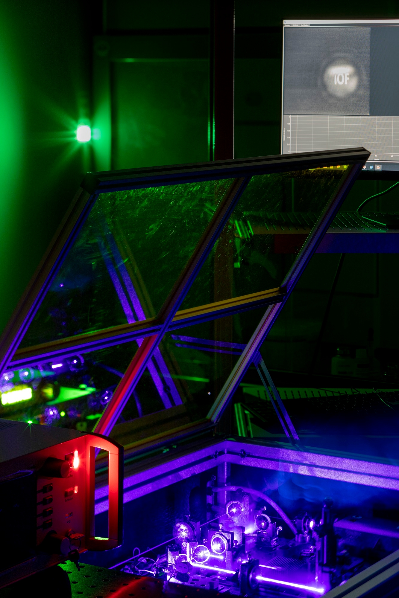
To analyze potential tumor tissue via quantum imaging, researchers from the Fraunhofer Institute for Applied Optics and Precision Engineering IOF in Jena, the Technical University of Darmstadt Germany, and the Barcelona Institute of Science and Technology use a nonlinear crystal. A laser beam is sent through it, which gives out correlated light particles, so called entangled photon pairs. One of these photons is send into the tissue sample that is supposed to be pictured, while the “twin” is send to the optical imaging sensor. So, while one photon illuminates the object, the camera only captures the twin. Through the entanglement of the particles, the information recorded from the tissue is transferred to the photons detected by the camera and made visible, which is how an image of the object is produced.
But one problem has persisted up to now: Quantum imaging is susceptible to the interference of surrounding light and must therefore be conducted in an isolated environment. This has limited the application of the process in practice so far. Now, the researchers have perfected a process to the point surrounding light, or so called “optical noise”, has hardly any effect on quantum imaging.
Quantum imaging distillation with undetected light
In an experiment the team of researchers were able to clean a quantum image from noise by means of a distillation technique. As opposed to methods applied so far, which use the joint detection of photon pairs, the researchers introduce a quantum imaging distillation technique that uses the detection of single photons only. This imaging distillation approach is based on the interferometric modulation of the interfering signal. Here, the original signal is varied in brightness, while the optical noise changes very little over time. This makes it possible to separate both signals from each other and remove the interfering light.
The researchers were able to prove the effectiveness of the process by projecting a noise image, i.e., an unwanted signal, in addition to the quantum image on the image sensor and subsequentially were able to remove it successfully. Even with interfering light that is up to 250 times more intense than the light that produced the image, the method works. The researchers estimate based on their calculations that even an interfering light that is up to 1,000 times more intense can be removed successfully.
New tool for biomedicine
The new process represents a significant advance in quantum imaging. At Fraunhofer IOF a quantum imaging system is now supposed to be transferred into a model for use in the field. The main goal of the project is to create a highly developed quantum imaging system that can be integrated seamlessly into a scanning microscope. This leading technology is a crucial step towards revolutionizing the imaging process in various areas, such as medicine and materials sciences.
The process of quantum imaging distillation with undetected light, according to Prof. Markus Gräfe of the Technical University of Darmstadt, offers a promising “tool for biomedicine”. Here, infrared light is often used for sample analysis. But when the actual detection is carried out with visible light, the hope is that more information reaches the images. Although the idea of using quantum imaging in this field is not new, the novel process is characterized by the foregoing of elaborate shielding measures and could in this way be easily integrated into a commonplace laboratory environment.
The experiment took place within several research projects – among them QUILT, Multi-Use, SPECTRA, and the Quantum Hub Thuringia. The project QUANCER is currently continuing the work in this research area.
Original publication
Jorge Fuenzalida, Marta Gilaberte Basset, Sebastian Töpfer, Juan P. Torres, Markus Gräfe: »Experimental quantum imaging distillation with undetected light«, Science Advances, Vol. 9, No. 35: DOI: 10.1126, URL: https://www.science.org/doi/10.1126/sciadv.adg9573.
